© Brighteon.com All Rights Reserved. All content posted on this site is commentary or opinion and is protected under Free Speech. Brighteon is not responsible for comments and content uploaded by our users.
This is a drop of jabbed blood on a slide magnified 40x using a microscope. The red coloring you see is the actual red blood cells. The black is magnetographene and always appears powder-like in the blood. The light green structures with the dark black outline are Lipid Nano Gel structures and are full of other structures and substances when viewed at a higher magnification. What you see in the video is the MagnetoGraphene pushing and spreading the Lipid Nano Gel throughout the blood on the slide.
Check out the YouTube video: Nanotubes assemble! Rice introduces Teslaphoresis
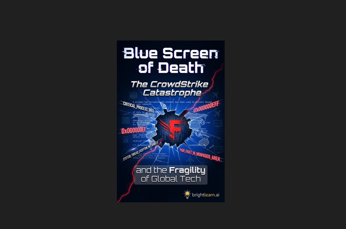
Blue Screen of Death: How CrowdStrike’s global meltdown exposed the deadly flaws of centralized control
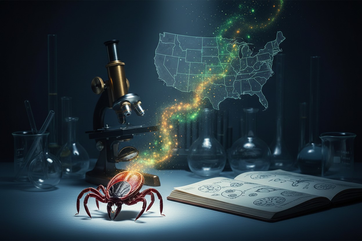
CIA’s Cold War Tick Experiments Examined Following New Allegations Linking Them to Lyme Outbreak

The longevity molecule: How a common amino acid could rewrite the rules of aging

Study Reviews Potential Immune System Effects of Green Tea Consumption

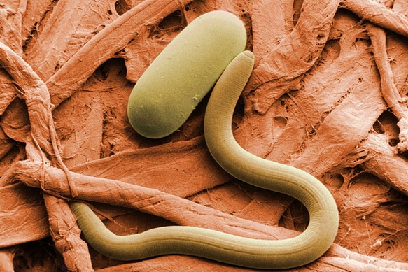
Declassified CIA files reveal Cold War-era Soviet research linking parasites to cancer treatment